BÖBREK TAŞ HASTALIKLARI
Kidney Stone Diseases
Channel D As We Speak - Kidney Stones - Assoc. Prof. Dr. Uygar Miçooğulları
Kidney Stone

What is the Function of the Kidneys?
The kidneys are vital organs responsible for filtering harmful waste products from the blood and eliminating them from the body in the form of urine, along with water. In addition to urine production, the kidneys perform several other critical functions.
One of their essential roles is the production of **erythropoietin**, a hormone that stimulates the maturation of red blood cells in the bone marrow, thereby helping to protect the body against anemia.
Furthermore, the kidneys contribute to the synthesis of **vitamin D**, which plays a key role in regulating calcium and phosphorus balance. This regulation is crucial for maintaining strong and healthy bones.
How Are Kidney Stones Formed?
The kidneys are vital organs responsible for filtering harmful waste products from the blood and eliminating them from the body in the form of urine, along with water. In addition to urine production, the kidneys perform several other critical functions.
One of their essential roles is the production of **erythropoietin**, a hormone that stimulates the maturation of red blood cells in the bone marrow, thereby helping to protect the body against anemia.
Furthermore, the kidneys contribute to the synthesis of **vitamin D**, which plays a key role in regulating calcium and phosphorus balance. This regulation is crucial for maintaining strong and healthy bones.
How Many Types of Kidney Stones Are There?
Taşlar yapısını oluşturan kristallerin cinsine göre 5 ana kategoriye ayrılmaktadır: Kalsiyum Oksalat Taşları (en sık), Kalsiyum Fosfat Taşları, Enfeksiyon (Strüvit) Taşları, Ürik Asit Taşları ve Sistin Taşları. Taşın yapısının bilinmesi, tedavi sonrası tekrar taş oluşumunun önlenmesi ve buna yönelik önlemler alınması açısından önemlidir.


What are the Symptoms of Kidney Stone Disease?
Kidney Stone Disease
Kidney stones can sometimes be detected incidentally without causing any symptoms; however, they may also migrate into the urinary tract, leading to severe pain. The nature of the pain is typically **colicky**, meaning it comes in waves and can be excruciating. The pain is usually felt in the **flank region** and may radiate towards the groin area.
In addition to pain, the most common symptoms include:
- **Nausea and vomiting**
- **Burning sensation during urination**
- **Presence of blood in the urine (hematuria)**
- **Difficulties with urination**
These symptoms can significantly affect daily life and may require medical intervention.
How is Kidney Stone Disease Diagnosed?
Diagnosis of Kidney Stone Disease
The diagnosis of kidney stone disease involves a thorough **medical history** and **physical examination**, along with various laboratory and imaging techniques. The most commonly used radiological imaging methods for detecting kidney stones include:
- **Ultrasonography**,
- **Direct X-ray (KUB - Kidney, Ureter, Bladder radiograph)**,
- **Non-contrast low-dose computed tomography (CT scan)**.
If an intervention is planned for the stone, **contrast-enhanced imaging** or, when necessary, **nuclear scintigraphy** may be performed for further evaluation.
To investigate the underlying causes of kidney stones, a series of **metabolic tests** on blood and urine, as well as **stone analysis**, are crucial. These tests play a significant role in preventing recurrence of the disease in the future.
Treatment of Kidney Stones
Treatment of Kidney Stone Disease
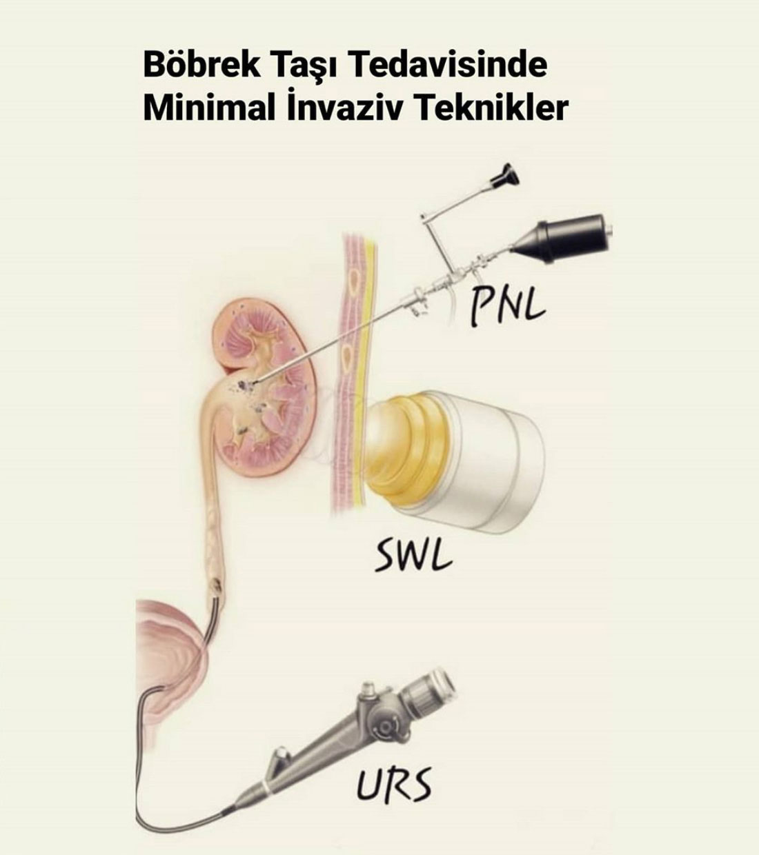
The treatment of kidney stone disease is planned based on several factors, including the **location, size, and type of the stone**, as well as individual patient characteristics. The primary treatment methods include:
- **ESWL (Extracorporeal Shock Wave Lithotripsy):** Breaking stones using sound waves.
- **URS / RIRS (Ureterorenoscopy / Retrograde Intrarenal Surgery):** Endoscopic procedures to remove or break stones in the urinary tract.
- **PNL (Percutaneous Nephrolithotomy):** A minimally invasive procedure to remove larger kidney stones through a small incision in the back.
- **Open and Laparoscopic Surgery:** Used in complex cases where other methods are not suitable.
To prevent the recurrence of kidney stones, patients are advised to adopt **lifestyle modifications, dietary changes**, and, based on metabolic evaluation results, **medication therapy** may be recommended.
Kidney Stone Breaking with Laser
Laser Lithotripsy
Laser lithotripsy is a procedure performed using a **flexible, thin endoscope** that allows access to the kidney through the urinary tract without the need for any incisions or punctures. The stones within the kidney are fragmented or removed with the assistance of a laser.
For this procedure, specially designed endoscopes known as **flexible ureterorenoscopes** are utilized. These instruments are approximately **3 mm in diameter** and **60–70 cm in length**, with a flexible tip that can be controlled by the surgeon from the handle of the device.
This advanced flexibility allows the endoscope to navigate through all chambers of the kidney, enabling precise targeting and fragmentation of stones using laser energy.
Kidney Stones in Infants and Children
**Kidney Stone Disease in Infants and Children**
Although kidney stone disease is less common in infants and children compared to adults, its incidence has been increasing in recent years due to changes in dietary habits. Additionally, pediatric kidney stones are often associated with underlying **metabolic or anatomical disorders**, making them highly prone to recurrence.
Due to the smaller size of children's kidneys and urinary tracts, the treatment of pediatric kidney stones requires the use of **miniaturized, specialized instruments**, and the surgeon must have substantial experience in this field.
I have one of the **largest case series in the world** regarding pediatric kidney stone surgery, with my **success rates exceeding the global literature standards**. I have published **over 80 international articles** on this topic. Throughout my career, I have provided training to numerous colleagues from both Turkey and abroad in the institutions I have worked with, organizing **dozens of symposiums and conferences** on pediatric stone surgery.
In 2011, we were the **first in the world** to publish a series on **minimally invasive laser kidney stone surgery in preschool children** (*Journal of Pediatric Surgery, 2011*), and at that time, we reported the youngest children ever treated with this surgical approach in the literature.
With all this experience and knowledge, I continue to perform pediatric kidney stone surgeries with **high efficiency and safety**, ensuring optimal outcomes for infants and children.
Breaking with Sound Waves (ESWL)

ESWL (Extracorporeal Shock Wave Lithotripsy)
In this technique, sound waves produced by a generator are transmitted to the kidney stone through the skin under fluoroscopy and/or ultrasound guidance, and the stone fragments broken by strong vibrations are expelled from the body through urine. The procedure takes approximately 30-45 minutes, depending on the size and location of the stone and the device used. Additional sessions may be needed to ensure complete stone-free operation, but it is definitely not recommended to have more than 3 sessions. The procedure is performed on an outpatient basis and there is no need to stay in the hospital afterwards. Thanks to new generation devices, there is no need for anesthesia except for pediatric patients. However, it is beneficial for the patient to use painkillers before the procedure so that they do not feel pain.
Who Can Get the ESWL Procedure?
**Factors Affecting the Success of the Procedure**
The success of the procedure depends on several factors, including:
- **Stone size, number, and location**,
- **Stone hardness**,
- **Anatomical structure of the kidney**,
- **Distance between the stone and the skin**.
This procedure is generally preferred for stones **smaller than 2 cm**. It can also be safely performed in **pediatric patients**, where stones tend to break down more easily and pass more smoothly compared to adults.
The only concern in pediatric cases is that the procedure must be performed **under anesthesia**, which requires careful monitoring and specialized pediatric anesthetic expertise to ensure safety.
Who Can't Get the ESWL Procedure?
**It is risky and inappropriate to perform this procedure in:**
- **Pregnant women**, due to potential risks to the fetus.
- Individuals **taking blood thinners** or those with **bleeding disorders**, as they have a higher risk of bleeding complications.
- Patients with **active urinary tract infections**, which may lead to severe infections or sepsis.
- Individuals with **aneurysms in major arteries**, as the procedure may exacerbate the condition and pose life-threatening risks.
- Those with **renal outlet obstruction**, which can hinder stone passage and complicate treatment outcomes.
In such cases, alternative treatment options should be carefully considered.
What are the advantages and disadvantages compared to surgical methods?
**The main advantages** of this procedure include:
- It **does not require anesthesia**, making it a safer option for certain patients.
- It has **fewer complications**, reducing the risk of unwanted side effects.
- It can be performed as an **outpatient, same-day procedure**, allowing patients to return to their daily activities quickly.
**However, the disadvantages** include:
- **Lower success rates**, particularly in cases involving **hard stones** or stones located in the lower calyx of the kidney.
- **Stone fragments** may cause **blockage or pain** as they pass through the urinary tract.
**Note:** The content on this page is for informational purposes only. Always consult your doctor for diagnosis and treatment.
Ureteroscopy(URS-RIRS)
It is an endoscopic (closed) surgical method performed by entering through the urinary tract, used in the treatment of ureteral stones (the thin canal that connects the kidney and bladder).


How To?
It is performed under general or spinal anesthesia. Rigid or flexible endoscopes can be used during the surgery and the procedure is named according to the device used. The patient is placed on his back and placed in the lithotomy (birth) position. The stone is broken into very small pieces with a laser stone breaking device sent through the endoscope. If necessary, stone pieces can be removed with the help of tools called basket catheters. If the stone is very large, has caused serious edema in the canal wall or has an injury, a stent can be placed between the kidney and bladder to allow urine flow. This stent is removed after approximately 1-2 weeks.
What are the advantages?
Since no incision or puncture is made, the patient can be discharged on the same day and can return to normal daily life the next day. It is an effective and safe surgical method with a very high success rate and a very low complication (unwanted side effect) rate. It can be safely applied to all patients except those with active urinary tract infections.
Are There Any Risks?
Although it is a closed (endoscopic) procedure, it does carry some risks. If the urinary tract is too narrow for the endoscope to pass, the procedure can be postponed to a second session by placing a stent. The most feared complication of the procedure is injury or rupture of the tract. While most of these injuries heal with a stent, surgical repair is required in cases of rupture or very serious injuries. Other rare complications that can be observed are infection, fever and bleeding, which require antibiotic treatment after surgery. Careful pre-surgical preparation, use of fine instruments and surgical experience are important to prevent such unwanted side effects.
Retrograde Intrarenal Surgery (RIRS)
It is an endoscopic (closed) surgical method performed by entering through the urinary tract, used in the treatment of ureteral stones (the thin canal that connects the kidney and bladder).

Laser Lithotripsy
Laser lithotripsy is a procedure performed using a **flexible, thin endoscope** that allows access to the kidney through the urinary tract without the need for any incisions or punctures. The stones within the kidney are fragmented or removed with the assistance of a laser.
For this procedure, specially designed endoscopes known as **flexible ureterorenoscopes** are utilized. These instruments are approximately **3 mm in diameter** and **60–70 cm in length**, with a flexible tip that can be controlled by the surgeon from the handle of the device.
This advanced flexibility allows the endoscope to navigate through all chambers of the kidney, enabling precise targeting and fragmentation of stones using laser energy.
What are the advantages?
Since no incision or puncture is made, the patient can be discharged on the same day and can return to normal daily life the next day. It is an effective and safe surgical method with a very high success rate and a very low complication (unwanted side effect) rate. It can be safely applied to all patients except those with active urinary tract infections.
Who Can Get the ESWL Procedure?
Although it is generally a preferred method for stones smaller than 2 cm, my personal experience and studies published in recent years indicate that this procedure can be successfully applied to stones up to 3 cm located outside the lower chamber of the kidney.
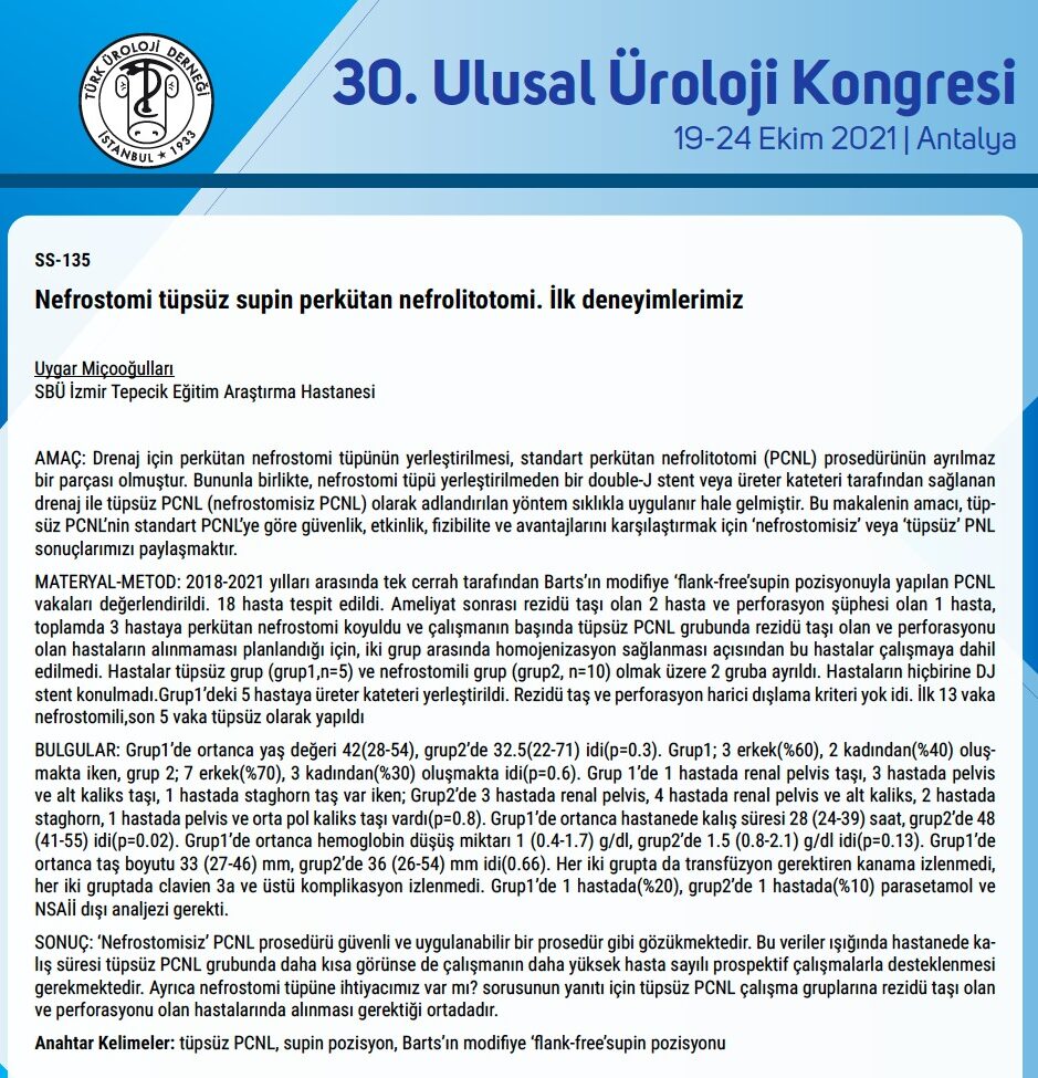
Perkütan Nefrolitotomi (PNL)
Perkütan Nefrolitotomi (PNL) böbrek taşlarının tedavisinde kullanılan, sırttan küçük bir delik açılarak gerçekleştirilen endoskopik (kapalı) bir ameliyat tekniğidir. Bu yöntem daha çok 2 cm’den daha büyük taşlarda veya diğer tedavi seçeneklerinin (ESWL ve RİRC) başarısız olduğu daha küçük böbrek taşlarında tercih edilir.
How To?
A tunnel is created by inserting a tube approximately 1 cm wide from the patient's back into the kidney. The stones inside the kidney are reached with the endoscope sent through this tunnel. These stones are removed as a whole or by breaking them into pieces with various crushers (pneumatic, ultrasonic or laser) and removed through the same path. If necessary after the intervention, a thin plastic tube (nephrostomy tube) is inserted into the kidney through the hole where the surgery was performed and is kept for 1-2 days for rapid recovery after the surgery. In small stones and surgeries that are completed without any problems, it is often not necessary to insert this tube.
Are There Different Types of This Surgery?
PNL surgery has become possible to perform through smaller incisions thanks to the thinner endoscopes and instruments developed in recent years. The surgery is named according to the diameter of the inserted tube; while a 30 Fr (1 cm) diameter tube is inserted in the standard technique, 15-18 Fr diameter tubes (5-6 mm) are used in the mini-PNL method, and 12-14 Fr diameter tubes (3-4 mm) are used in the ultramini-PNL technique. As the diameter of the tube used decreases, the possibility of some undesirable conditions such as bleeding due to surgery decreases. Again, recovery is faster after surgeries performed with smaller diameter instruments, the patient experiences less pain and has a more comfortable recovery process.
What are the advantages?
The most important advantage of this technique over other methods is that it is the method with the "highest chance of success (stone-free)" for stones larger than 2 cm. It provides a great advantage in terms of cosmetics and a faster and more comfortable recovery process compared to open stone surgeries, which were frequently used in the past for the treatment of large kidney stones and were performed through a large incision.
Are There Any Risks?
Although this surgery is considered a closed endoscopic method, it carries some risks such as bleeding, infection and injury to adjacent organs. The way to minimize these risks is to make the right preparation before the surgery, to have fully equipped equipment and to have sufficient experience of the surgeon in this surgery.
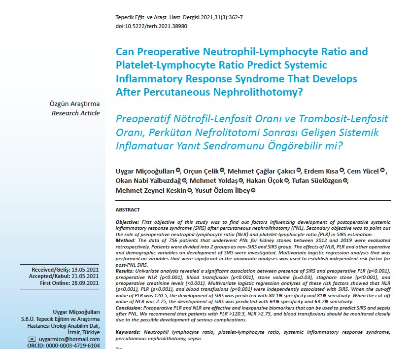
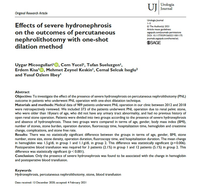
Some of My Studies on Percutaneous Nephrolithotomy