
KIDNEY STONE DISEASES
Assoc. Prof. Dr. Uygar Miçooğulları, FEBU · İzmir Urology and Andrology
How big is your stone?
If your report says "8 mm", see what that means with a familiar comparison from the kitchen. Move the slider to explore the chance of passing the stone by size and the treatments usually considered.
The chance of a stone passing depends not only on its size but also on its location (kidney/ureter), its composition and the anatomy of the urinary tract. Fever, persistent pain or inability to urinate are emergencies.
Lentil ≈ 2–4 mm
Small stone. Most ureteral stones under 5 mm pass on their own with plenty of fluids and pain control, usually within a few weeks.
Chickpea ≈ 5–9 mm
The chance of passing on its own decreases. Medication to help the stone pass may be tried; if it does not pass, or if there is pain, infection or kidney swelling, it is broken up with a laser by ureteroscopy (URS).
Kidney stone treatmentsHazelnut ≈ 10–20 mm
Usually does not pass on its own. In the kidney, minimally invasive laser surgery with flexible ureteroscopy (RIRS) or shock wave lithotripsy (ESWL); in the ureter, URS is often preferred.
Minimally invasive laser stone surgeryOlive > 20 mm
Large stone. For kidney stones over 2 cm, percutaneous nephrolithotomy (PCNL) is usually the first option; RIRS may be used in selected patients.
Kidney stone treatmentsStone Diseases
Kanal D Konuştukça - Kidney Stones - Op. Dr. Uygar Miçooğulları
Kidney Stones
What Is the Function of the Kidneys?
What Is the Function of the Kidneys?
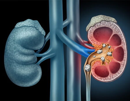
The kidneys are organs that filter harmful waste from the blood and allow this waste to be excreted from the body together with water in the form of urine. Apart from producing urine, the kidneys have other important functions. The kidneys are responsible for producing a hormone called erythropoietin, which matures the blood cells in the bone marrow. In this way, they protect the body against anemia. In addition, they regulate the calcium and phosphorus balance by contributing to the synthesis of vitamin D. This keeps our bones strong.
How Do Kidney Stones Form?
How Many Types of Kidney Stones Are There?
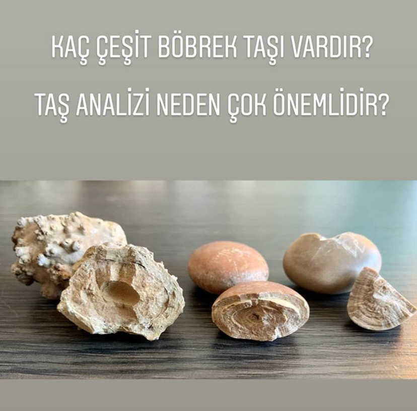
Stones are divided into 5 main categories according to the type of crystals that make up their structure: calcium oxalate stones (the most common), calcium phosphate stones, infection (struvite) stones, uric acid stones and cystine stones. Knowing the composition of the stone is important for preventing new stone formation after treatment and for taking measures to that end.

What Are the Symptoms of Kidney Stone Disease?
Kidney stone disease can sometimes be detected incidentally without causing any symptoms, while at other times stones can pass into the urinary tract and cause very severe pain. The pain is usually colicky in nature, coming in waves and making the person writhe. The pain is felt in the flank area and radiates toward the groin. Apart from pain, the most important symptoms are nausea, vomiting, burning during urination, blood in the urine and urination problems.
How Is Kidney Stone Disease Diagnosed?
In addition to a detailed medical history and examination, a number of laboratory and imaging methods are used to diagnose kidney stone disease. Ultrasonography, plain X-ray and low-dose non-contrast computed tomography are the most important radiological imaging methods used in stone diagnosis. When an intervention for the stone is planned, contrast-enhanced studies or, when necessary, scintigraphic studies may be planned. To investigate the causes of stone disease, carrying out certain metabolic tests on blood and urine and analyzing the stone are important for preventing the disease from recurring in the future.
Treatment of Kidney Stones
The treatment of kidney stone disease is planned by taking into account the location, size and type of the stone, as well as a number of patient-related factors.
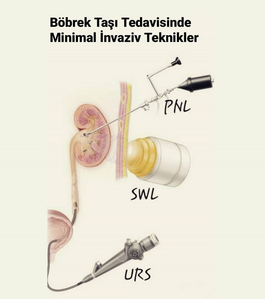
-ESWL (shock wave lithotripsy),
-URS / RIRS (ureterorenoscopy),
-PCNL (percutaneous nephrolithotomy),
-Open and laparoscopic surgical methods
are the treatment methods used for this purpose. Also, to prevent stone disease from recurring, patients may be advised to make certain lifestyle changes, follow dietary treatment and, depending on the results of metabolic investigations, take medication.
Laser Kidney Stone Treatment
Laser stone treatment is a procedure in which a thin, flexible endoscope is passed through the urinary tract up to the kidney without making any incision or hole in the body, and the stones there are broken up with a laser or removed. Specially manufactured endoscopes (flexible ureterorenoscopes) are used for laser kidney stone treatment. The tip of these instruments, which are approximately 3 mm thick and 60-70 cm long, can be steered by the surgeon from the back of the device. This makes it possible to enter each calyx of the kidney, reach the stones there and break them up with a laser.
Kidney Stones in Infants and Children
Although stone disease is rarer in infants and children than in adults, its incidence has been increasing in recent years due to changes in eating habits. In addition, because stone disease in these children often has an underlying metabolic or anatomical disorder, it is highly prone to recurrence. Because a child's kidney and urinary tract are small, special miniature devices must be used in treating this age group, and the surgeon must have sufficient experience in this field. I have one of the largest series in the world in kidney stone surgery in children, and my success rates are well above the general literature. I have published more than 80 international articles on this subject. At the institutions where I have worked, we have trained many colleagues from Turkey and abroad in these surgeries. We have organized dozens of symposia and conferences. We were the first in the world to publish a series of laser minimally invasive kidney stone surgery in preschool children (J Ped Surg 2011). At that time, we were the first to report in the literature the youngest children on whom these surgeries had been performed. With all this experience and knowledge, I continue to perform stone surgeries in infants and children very frequently, effectively and safely.
Shock Wave Lithotripsy (ESWL)
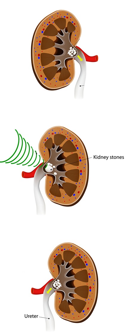
ESWL (Extracorporeal Shock Wave Lithotripsy)
In this technique, shock waves produced by a generator are transmitted through the skin to the kidney stone under fluoroscopy and/or ultrasound guidance, and the stone fragments broken up by the powerful vibrations are expelled from the body through the urine. The procedure takes approximately 30-45 minutes, varying according to the size and location of the stone and the device used. Additional sessions may be needed to achieve complete stone clearance, but more than 3 sessions are definitely not recommended. The procedure is performed on an outpatient basis and there is no need to stay in hospital afterward. Thanks to new-generation devices, anesthesia is not required except for pediatric patients. However, it is beneficial for the patient to take a painkiller before the procedure so as not to feel pain.
Who Is ESWL Suitable For?
Although the success of the procedure depends on many factors such as the size, number, location and hardness of the stone, the anatomical structure of the kidney and the distance between the stone and the skin, it is generally preferred for stones smaller than 2 cm. This procedure can also be applied safely in pediatric patients; in fact, in children stones are broken more easily with this method and pass more easily. The only drawback for pediatric patients is that the procedure is performed under anesthesia.
Who Is ESWL Not Suitable For?
Performing this procedure is risky and inadvisable in pregnant women, in those taking blood-thinning medication or with bleeding disorders, in those with an active urinary tract infection, in those with an aneurysm of the main arteries and in those with ureteropelvic junction (UPJ) obstruction.
What Are Its Advantages and Disadvantages Compared with Surgical Methods?
Its most important advantages are that it does not require anesthesia, has a lower rate of adverse effects (complications) and can be performed as a same-day outpatient procedure. Its disadvantages are a lower success rate (especially for hard stones and stones located in the lower calyx of the kidney) and the possibility that stone fragments may cause obstruction or pain as they pass.
NOTE: The content of this page is for informational purposes only; always consult your doctor for diagnosis and treatment.
Ureteroscopy (URS)
It is an endoscopic (minimally invasive) surgical method performed by entering through the urinary tract, used in the treatment of ureteral stones (the ureter is the thin tube connecting the kidney and the bladder).

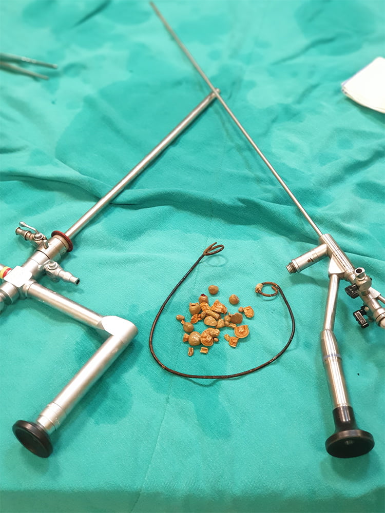
How Is It Performed?
It is performed under general or spinal anesthesia. Rigid or flexible endoscopes can be used during the operation, and the procedure is named according to the device used. The patient lies on their back and is placed in the lithotomy position. The stone is broken into very small fragments with a laser lithotripsy device passed through the endoscope. When necessary, stone fragments can be removed with instruments called basket catheters. If the stone is very large, has caused significant edema in the ureteral wall or an injury has occurred, a stent may be placed between the kidney and the bladder to ensure urine flow. This stent is removed after approximately 1-2 weeks.
What Are the Advantages?
Since no incision or puncture is made, the patient can be discharged on the same day and return to normal daily life the next day. It is an effective and safe surgical method with a very high chance of success and a very low rate of complications (adverse side effects). It can be applied safely to all patients except those with an active urinary tract infection.
Are There Any Risks?
Although it is a minimally invasive (endoscopic) procedure, it carries some risks. If the ureter is too narrow for the endoscope to pass, a stent may be placed and the procedure postponed to a second session. The most feared complication of the procedure is injury to or avulsion of the ureter. While most of these injuries heal with a stent, surgical repair is required in cases of avulsion or very serious injury. Infection requiring antibiotic treatment after surgery, fever and bleeding are other rare complications. To prevent such adverse effects, careful preoperative preparation, the use of fine instruments and surgical experience are important.
Retrograde Intrarenal Surgery (RIRS)
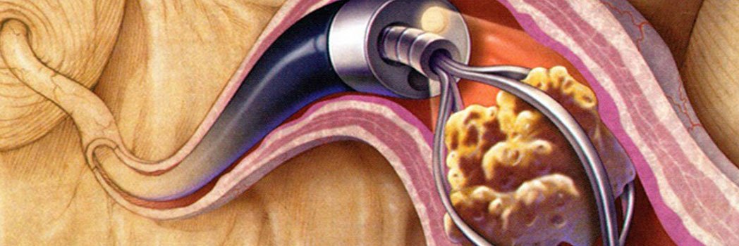
It is a procedure in which a thin, flexible endoscope is passed through the urinary tract up to the kidney without making any incision or hole in the body, and the stones there are broken up with a laser or removed. Specially manufactured endoscopes (flexible ureterorenoscopes) are used for this procedure. The tip of these instruments, which are approximately 3 mm thick and 60-70 cm long, can be steered by the surgeon from the back of the device. This makes it possible to enter each calyx of the kidney, reach the stones there and break them up with a laser.
Since no incision or puncture is made, the patient can be discharged on the same day and return to normal daily life the next day. It is a surgical technique with a very high chance of success and a low rate of complications (adverse side effects). It can be applied safely to all patients except those with an active urinary tract infection.
Who Is RIRS Suitable For?
Although it is generally a method preferred for stones smaller than 2 cm, my personal experience and studies published in recent years indicate that this procedure can be applied successfully for stones up to 3 cm located outside the lower calyx of the kidney.
Percutaneous Nephrolithotomy (PCNL)
Percutaneous nephrolithotomy (PCNL) is an endoscopic (minimally invasive) surgical technique used in the treatment of kidney stones, performed through a small opening made in the back. This method is mostly preferred for stones larger than 2 cm or for smaller kidney stones for which other treatment options (ESWL and RIRS) have failed.
A tunnel is created by placing a tube approximately 1 cm wide from the patient's back into the kidney. The stones inside the kidney are reached with an endoscope passed through this tunnel. These stones are removed through the same route either whole or after being fragmented with various lithotripters (pneumatic, ultrasonic or laser). After the procedure, if necessary, a thin plastic tube (nephrostomy tube) is placed into the kidney through the opening used for the operation and left for 1-2 days for rapid postoperative recovery. For small stones and operations completed without problems, it is often not necessary to place this tube.
Are There Different Types of This Operation?
Thanks to thinner endoscopes and instruments developed in recent years, PCNL can now be performed through smaller incisions. The operation is named according to the diameter of the tube placed: in the standard technique a tube 30 Fr (1 cm) in diameter is placed, while in the mini-PCNL method tubes 15-18 Fr (5-6 mm) in diameter are used, and in the ultra-mini-PCNL technique tubes 12-14 Fr (3-4 mm) in diameter are used. As the diameter of the tube decreases, the likelihood of certain undesirable events such as surgery-related bleeding decreases. In addition, after operations performed with smaller-diameter instruments, recovery is faster, the patient feels less pain and experiences a more comfortable recovery process.
The most important advantage of this technique over other methods is that it is the method with the “highest chance of success (stone clearance)” for stones larger than 2 cm. Compared with open stone surgery performed through a large incision, which was frequently used in the past to treat large kidney stones, it offers a major cosmetic advantage and a faster, more comfortable recovery process.
Although this operation is considered a minimally invasive endoscopic method, it carries some risks such as bleeding, infection and injury to neighboring organs. The way to minimize these risks is correct preoperative preparation, fully equipped facilities and a surgeon with sufficient experience in this operation.
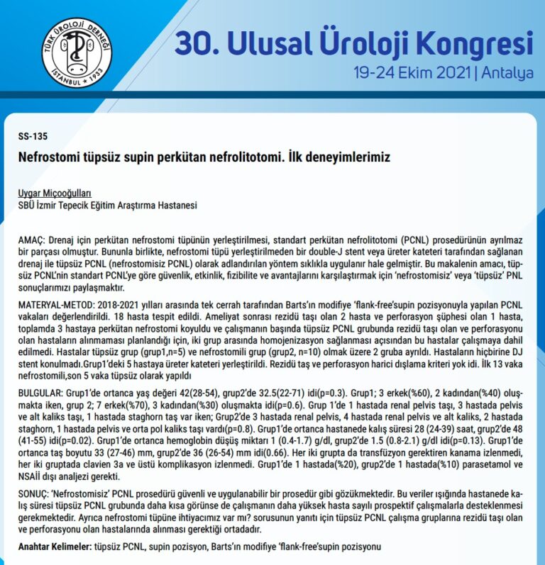
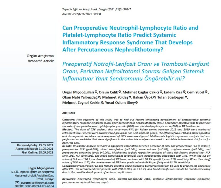
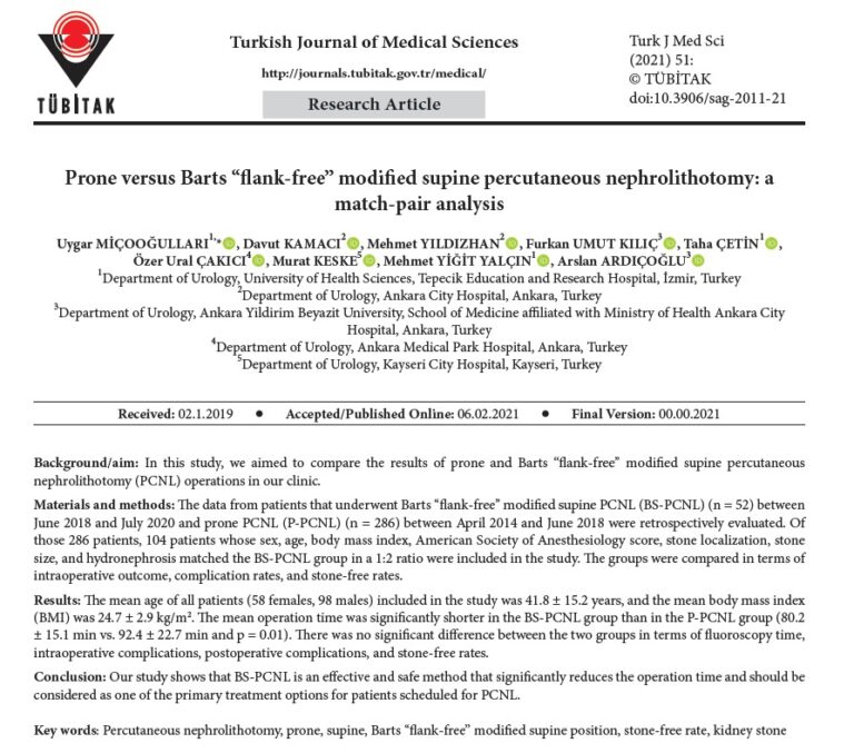
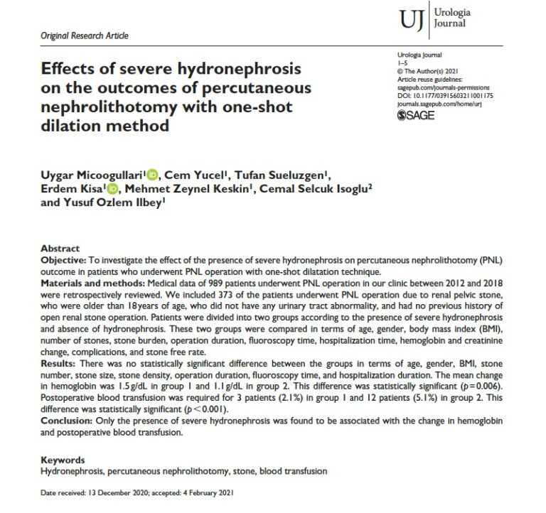
Some of My Studies on Percutaneous Nephrolithotomy
Contact us for an examination and appointment
Medical Park İzmir Hospital · Appointments: 0530 574 79 89 · 444 44 84
Book an Appointment