
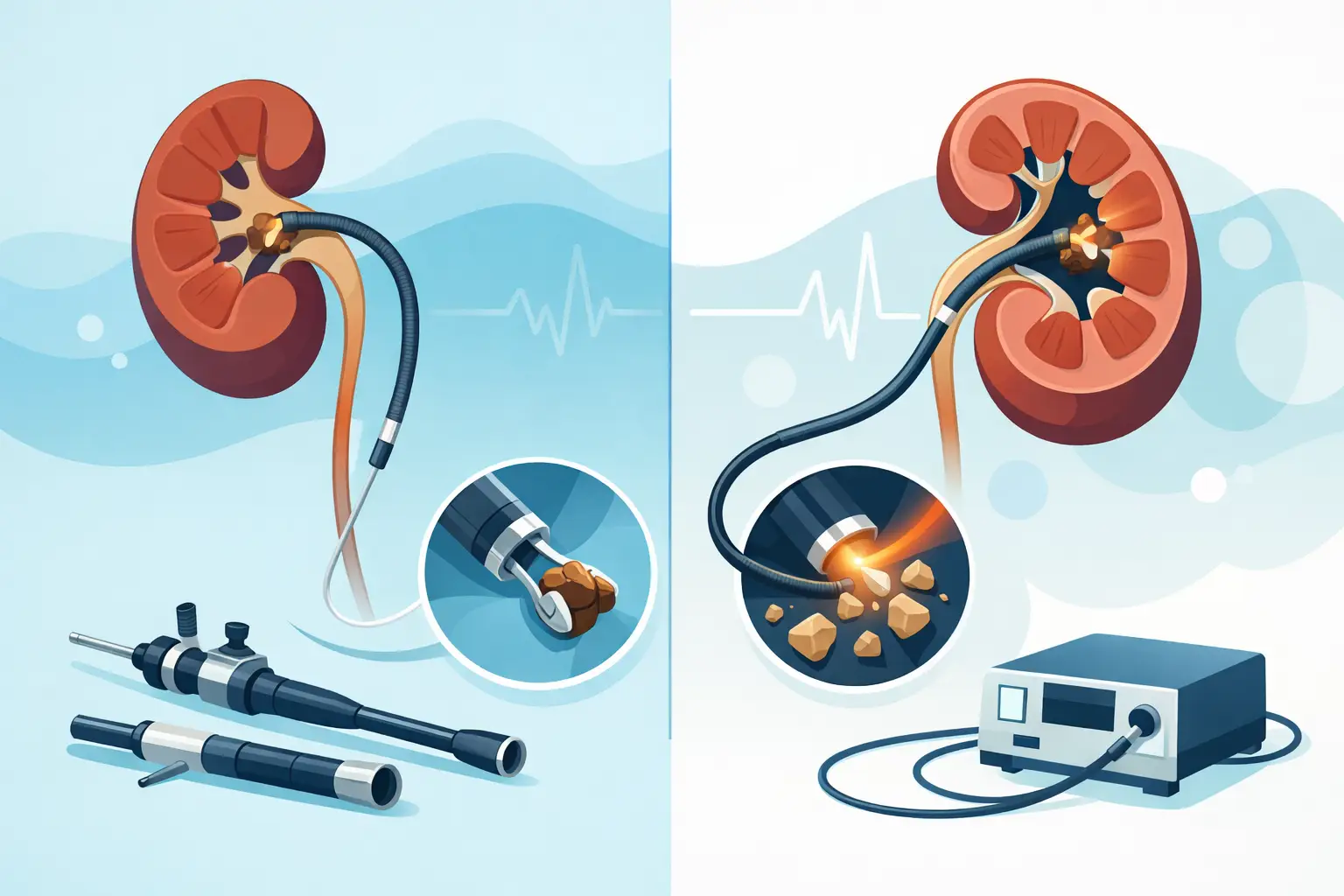
One of the most common questions asked by patients diagnosed with kidney stones is the difference between URS and RIRS and which method is more suitable for them. Both procedures are minimally invasive stone operations performed without any incision in the body, by advancing through the urinary tract. However, the flexibility of the instrument used, the area reached and, in particular, the location of the stone in the kidney or ureter change the choice of treatment.
The aim of stone treatment is not only to break up the stone. The goals are to clear the stone in the safest way possible, preserve kidney function, reduce the risk of infection and allow the patient to return to daily life in a controlled manner. For this reason, the decision between URS and RIRS is not made by looking only at the size of the stone in millimeters.
Where does the difference between URS and RIRS lie?
URS is the minimally invasive method called ureteroscopy. The ureter is the name given to the urinary channel between the kidney and the bladder. In URS, the bladder and then the ureter are reached by entering through the urinary channel. Thanks to the thin, semi-rigid endoscopes used especially for treating stones in the ureter, the stone is seen, broken up with a laser and, where appropriate, the fragments are removed.
RIRS is the abbreviation for retrograde intrarenal surgery. This method also uses the natural urinary passages; however, it uses a flexible instrument that can reach the chambers inside the kidney called calyces. With the flexible ureteroscope, it may be possible to reach stones in the upper, middle or lower part of the kidney with a wider range of movement.
In short, the basic distinction is this: URS is mostly referred to as an approach for ureteral stones, while RIRS is the flexible endoscopic technique used to reach stones inside the kidney. That said, in medical language the term URS can sometimes be used to cover all ureteroscopic procedures. For this reason, it is better for patients to learn clearly not the name of the procedure, but where the stone is located and the planned surgical approach.
When is URS preferred for a ureteral stone?
Ureteral stones can present with severe flank pain, nausea, vomiting, blood in the urine and sometimes infection. In particular, if the stone blocks urine flow, the kidney may become dilated. Some small stones may pass on their own; however, the size and location of the stone, pain that cannot be controlled and the effect on kidney function determine whether a procedure is needed.
URS is an effective option, particularly for stones in the middle and lower part of the ureter. The procedure is performed under general anesthesia. The surgeon sees the stone directly through camera systems and breaks it into small fragments with laser energy. Depending on the structure and location of the stone and the anatomical features of the ureter, the fragments may be removed or reduced to a size that can pass on its own.
For stones in the upper part of the ureter, close to the kidney, it may be necessary to proceed with flexible instruments instead of semi-rigid URS. At this point, the procedure may technically take on the characteristics of RIRS. When deciding, CT images and the anatomy of the urinary tract are evaluated in detail.
For which kidney stones does RIRS stand out?
RIRS is one of the minimally invasive methods that can be considered for many stones located inside the kidney, particularly those up to 2 centimeters. The steerable tip of the flexible instrument makes it easier to reach different chambers of the kidney. This feature can provide an advantage in selected lower calyx stones as well as in upper and middle calyx stones.
During RIRS, the stone can be reduced to dust with the laser or split into larger fragments. The surgical strategy is determined by the hardness of the stone and the total stone burden. With very hard stones, the procedure may take longer. When stones are present in more than one area, the stone burden is high or the kidney anatomy makes access difficult, becoming completely stone-free in a single session may not always be realistic. In some patients, planning a second session may be the safer and more appropriate approach.
For larger kidney stones, the PCNL method, known as percutaneous nephrolithotomy, may come into consideration. In PCNL, a small access is made into the kidney through the skin. For this reason, RIRS is not automatically the best method for every kidney stone. The most suitable option depends not only on the size of the stone but also on its location, density, the structure of the kidney and the patient's overall health.
What to expect during the procedure and recovery
Both URS and RIRS are mostly performed under general anesthesia. Before the procedure, a urinalysis and, when needed, a urine culture are evaluated. If there is an active urinary tract infection, it must be treated first. Computed tomography provides valuable information for surgical planning about the exact location, size and density of the stone and its relationship with surrounding tissues.
After surgery, a thin tube called a DJ stent is placed in some patients. This stent is used to support urine flow from the kidney to the bladder and to prevent swelling that may develop after the procedure from causing a blockage. A stent is not mandatory in every patient; however, the location of the stone, the duration of the procedure, sensitivity in the ureter and the likelihood of remaining fragments affect this decision.
While a DJ stent is in place, frequent urination, slight blood in the urine, discomfort in the bladder area or flank pain that may increase with movement can occur. These symptoms are usually temporary; however, in the case of high fever, chills, increasing pain, inability to urinate or heavy bleeding, you should contact your doctor without delay. The patient should be told clearly when the stent will be removed.
A large proportion of patients can return to daily life within a short time. Nevertheless, the pace of recovery varies from person to person. The size of the stone, the duration of the procedure, use of a stent, accompanying infection and the person's workload affect this process. In the first days, drinking enough fluids, taking the recommended medications regularly and following the follow-up plan are important.
URS or RIRS: Factors that determine the decision
When choosing the right method, not only the diameter of the stone but also the anatomical area where it is located is evaluated. An 8-millimeter stone in the lower ureter and an 8-millimeter stone in the lower calyx of the kidney may pose different technical challenges despite being the same size.
In the physician's evaluation, in addition to the size and location of the stone, stone density, intrarenal anatomy, previous surgery, the presence of infection, kidney function and any blood thinners used are taken into account. In patients with excess weight, skeletal problems or accompanying illnesses, the anesthesia and procedure plan is personalized separately.
The success of URS and RIRS depends on combining appropriate technology with experienced surgical planning. The type of laser, the quality of imaging, the features of the flexible instruments used and the management of stone fragments can affect the safety of the operation and the stone-free rate. That said, no method guarantees the same result in all patients. The aim is to choose the path that will provide the greatest clinical benefit with the least unnecessary procedural burden for the patient.
Possible risks of minimally invasive stone surgery
Although URS and RIRS are methods that can be performed with smaller interventions than open surgery, they are surgical procedures. Temporary burning on urination, bleeding, stent-related complaints and small stone fragments remaining in the kidney or ureter may occur. Rarely, infection, fever, injury to the ureter or the need for an additional procedure may arise.
These risks are minimized by controlling infection before the procedure, correct patient selection, use of appropriate equipment and close follow-up after surgery. Signs of infection such as fever and chills in particular should not be taken lightly. In stone disease, successful treatment is not completed when the operation ends, but with follow-up imaging and planning aimed at reducing new stone formation.
Clarifying during the examination whether your stone is in the kidney or the ureter, which result is aimed for in a single session and whether a stent will be needed makes the decision process easier. Assoc. Prof. Dr. Uygar Miçooğulları creates a personalized treatment plan for minimally invasive laser stone surgery by evaluating the characteristics of the stone and the patient's expectations together. Planning based on accurate information both reduces unnecessary anxiety and helps you start the recovery period better prepared.