Sudden, severe pain spreading from the lower back toward the groin raises the same question for many patients: will a kidney stone pass on its own? The answer depends not only on the presence of the stone but on its diameter, its position in the ureter, how well the kidney is draining urine, signs of infection and the patient's overall health. Some small stones can be passed naturally through the urinary tract, while others require treatment without waiting.
In stone disease, the right approach should not be limited to temporarily suppressing the pain. The aim is both to relieve the patient and to detect in time any situations that pose a risk of lasting kidney damage, infection and recurrent attacks.
Will a kidney stone pass on its own?
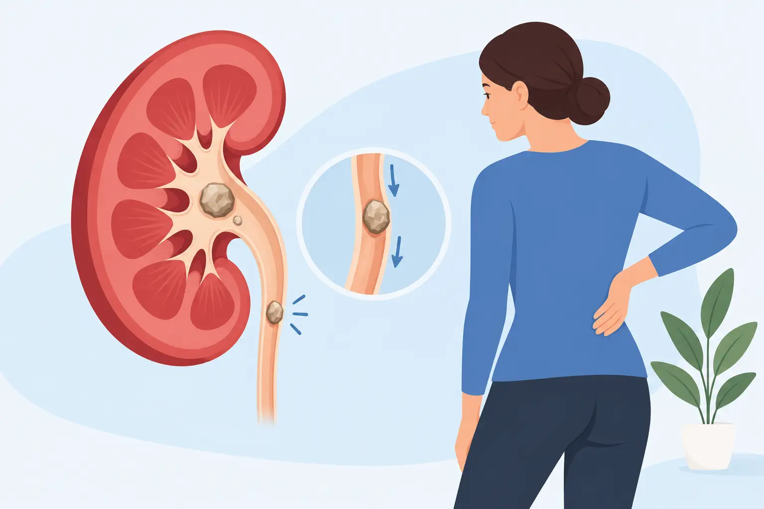
Not every stone inside the kidney is expected to pass on its own. For a stone to be passed with urine, it must first move from the kidney into the ureter, the thin tube between the kidney and the bladder. Whether the stone can progress through the ureter during this passage is assessed largely according to its diameter and its level.
In general, stones smaller than 5 millimeters in diameter are more likely to pass on their own. However, this does not mean that every small stone will definitely pass. A stone in the upper part of the ureter may progress with more difficulty than a stone of the same size near the lower part. For stones in the range of about 5-7 millimeters, the decision to wait is made more carefully; with larger stones, the likelihood of natural passage decreases markedly.
The shape of the stone also matters. Rough, sharp or impacted stones in the ureter can cause more pain and irritation. Having passed a stone before does not, on its own, indicate that a new stone will pass. Each stone attack should be evaluated separately with current imaging and examination findings.
Factors that affect stone passage
Stone size is one of the most decisive factors, but it is not the only criterion. Small stones located at the lower end of the ureter, close to the bladder, are generally more likely to pass naturally. In contrast, stones lodged at the kidney outlet or in the narrow parts of the ureter can cause prolonged pain and obstruction even if they are small.
Whether the stone causes urine to build up in the kidney is also critically important. This condition, described on imaging as hydronephrosis, suggests that urine cannot drain adequately from the kidney. Prolonged obstruction can adversely affect kidney function. Particularly in patients with a single kidney, in people with weak kidney function, or when obstruction on both sides is suspected, a waiting approach may not be appropriate.
The patient's level of pain and response to medication also change the treatment decision. If there is colic pain that stops daily life despite painkillers and requires frequent emergency visits, it is not right to focus only on the small size of the stone. The person's safety and quality of life are an integral part of the planning.
What can happen while a stone is passing?
As the stone moves within the ureter, flank pain coming in waves, pain radiating to the groin or testicle, nausea, vomiting and burning on urination may occur. A pink or red discoloration of the urine may also develop due to the stone irritating the wall of the urinary tract. Although these symptoms are common in stone disease, it is not possible to tell where the stone is or whether it has passed by looking at the symptoms alone.
In some patients, a sudden stop in pain gives the impression that the stone has been passed. However, the stone may have temporarily moved into a wider area, or the pain may have eased while the obstruction continues. For this reason, the stone should not be assumed to have passed completely without follow-up imaging.
In patients the physician considers suitable, medications that can help relax the ureter may be used. These medications are not necessary or safe for every patient; they should be planned taking into account blood pressure, other medications used and the position of the stone. Pain control and treatment for nausea can also help make the process more comfortable.
Does drinking plenty of water make the stone pass right away?
Adequate fluid intake is valuable for reducing stone formation and supporting urine flow. However, forcing yourself to drink a large amount of water during active, severe kidney stone pain does not make the stone pass immediately. In an obstructed system, it may even increase the pressure inside the kidney and make the pain more pronounced.
For this reason, fluid recommendations should be tailored to the individual. Patients who are vomiting, whose pain cannot be controlled or whose urine output is decreasing should seek medical evaluation rather than trying to drink liters of water on their own. Adjusting daily fluid habits after the stone has passed and the acute phase is over is more meaningful for preventing new stone formation.
When is urgent evaluation needed?
Stone pain is not always something that can be waited out at home. If any of the following symptoms are present, a same-day urological or emergency evaluation is needed:
- Fever, chills, marked weakness or deterioration in general condition
- Severe pain that continues despite painkillers
- Persistent vomiting and inability to keep fluids down
- Inability to pass urine or a marked decrease in the amount of urine
- Having a single kidney, pregnancy, known kidney failure or conditions affecting the immune system
In particular, the presence of an obstructing stone together with fever may indicate a urinary tract infection affecting the kidney under pressure. This situation requires prompt treatment. When necessary, drainage of the kidney is ensured first, the infection is brought under control and stone treatment is planned for a safe time.
Why do diagnosis and follow-up require imaging?
In urological evaluation, urinalysis, blood tests and imaging methods are used together. Ultrasound is useful for showing whether the kidney is dilated. Low-dose computed tomography can provide more detailed information for assessing the size, exact location and density of the stone, as well as alternative problems.
The key question in follow-up is not only "has the stone been seen?" It is assessed whether the stone is blocking the kidney's drainage, whether it is causing signs of infection and whether it is progressing within a certain period. This information makes it possible to distinguish patients who can safely wait from those who will benefit from a procedure.
What treatments are used if the stone does not pass?
If the likelihood of spontaneous passage is low, the pain keeps recurring, the obstruction persists or there is a risk of infection, minimally invasive treatments are considered. The method is chosen individually by evaluating the location, size and hardness of the stone, the kidney's anatomy and the patient's previous treatments.
For stones in the ureter, the stone can be reached with ureteroscopy (URS), entering the urinary channel with thin endoscopes. The stone can be broken into small fragments with laser energy and the fragments removed when necessary. For suitable stones inside the kidney, laser treatment using flexible endoscopes can be planned with retrograde intrarenal surgery (RIRS). For larger or more complex kidney stones, methods such as percutaneous nephrolithotomy (PCNL) may be preferred.
The purpose of these treatments is not only to clear the stone. The aims are to protect kidney function, reduce the risk of pain and infection and, where possible, avoid the burden of open surgery. In follow-up after the procedure, stone analysis, urine and blood evaluations, and dietary and fluid habits should also be addressed in a way that reduces new stone development.
Why should tests be done after the stone passes?
For someone who has passed a stone, the problem cannot always be considered completely over. Stone disease can recur, and the composition of the stone provides important information about why it formed. If possible, the passed stone should be kept in a clean container and brought to the physician for analysis.
In recurrent stones, a metabolic evaluation can assess calcium, citrate, uric acid and other risk factors in the urine. In some patients, reducing salt intake, increasing fluid intake or targeted dietary changes are sufficient; in others, medication may be needed. Applying the same "stone diet" to everyone is not appropriate, because stone types and causes differ.
Even if a kidney stone is small enough to wait for, it requires safe follow-up. The right path is determined by evaluating together the severity of your pain, the presence of fever, obstruction in the kidney and the stone's characteristics on imaging. Seeing a urologist without delay when in doubt reduces both unnecessary waiting and preventable complications.