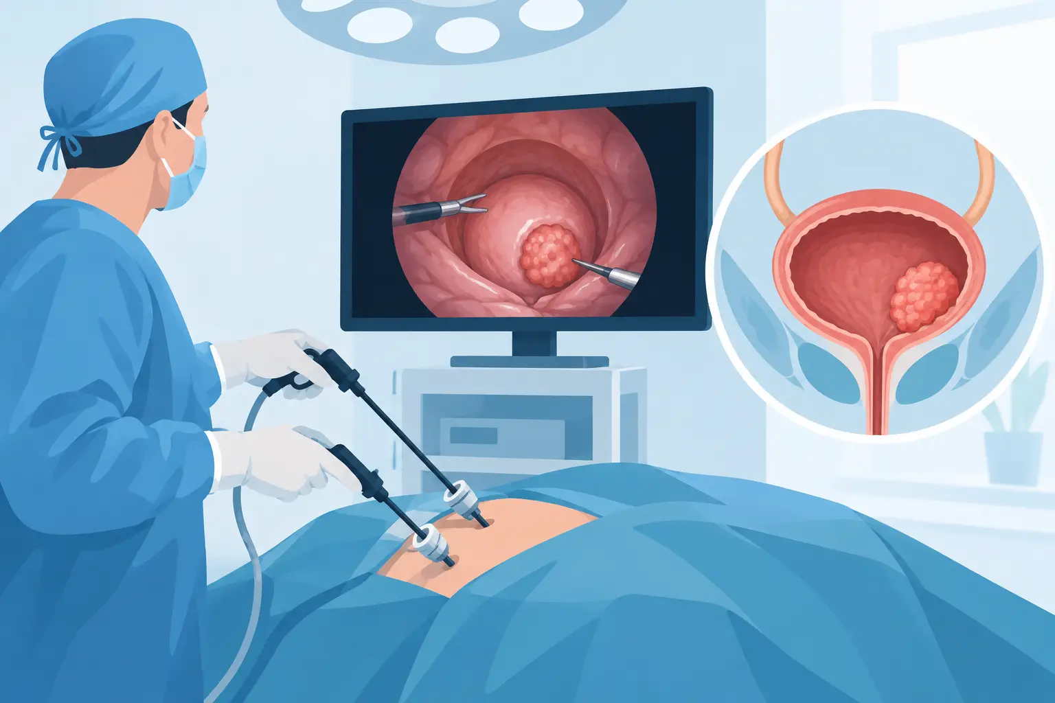
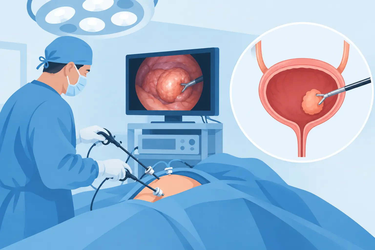
One of the most critical first steps in diagnosing bladder cancer is not only visualizing the tumor but removing it with the right method and evaluating it pathologically. Transurethral resection of bladder tumor, or TURBT, commonly known as minimally invasive bladder cancer surgery, is a procedure performed through the urethra that does not require an abdominal incision. The aim of this surgery is to remove the tumor as completely as possible, determine the stage of the disease and plan subsequent treatment individually.
A single surgical approach is not suitable for everyone with bladder tumors. The number and size of the tumors, how far they have progressed into the bladder wall, the pathology result, whether they have recurred before and the patient's general health are evaluated together. For this reason, while minimally invasive surgery is the main step of both diagnosis and treatment in some patients, in others it provides the information needed to move on to more advanced treatments.
What is minimally invasive bladder cancer surgery?
TURBT is the procedure of examining the inner surface of the bladder with a special camera system advanced through the urethra and removing the detected tumor with the help of a thin loop that uses electrical energy. No incision is made in the abdomen during the procedure. The surgeon sends the tumor tissue and the muscle tissue at the base of the tumor to pathology as separate samples.
The presence of muscle tissue in the sample is important. This is because, through this examination, the pathologist evaluates whether the tumor is confined to the inner surface of the bladder or has progressed into the bladder muscle. This is one of the key pieces of information that determines the scope of treatment.
The term minimally invasive should not lead one to think the procedure is simple. In some areas of the bladder, particularly for tumors near the openings of the ureters into the bladder or near the bladder neck, surgical experience is especially important. The goal is to clear the tumor to an adequate depth while preserving bladder function and the safety of surrounding structures.
In which patients is it performed?
When a tumor is suspected within the bladder, TURBT is usually the first-choice procedure. It comes into consideration in cases of visible blood in the urine, recurrent urine with clots, a mass detected in the bladder on imaging, or a suspicious area seen on cystoscopy.
Particularly in bladder cancers that have not progressed into the muscle layer, minimally invasive surgery can be directly curative in terms of completely removing the tumor. On the other hand, in disease that has progressed into the muscle layer, TURBT is still indispensable; but it generally does not mean it is sufficient on its own as the final treatment. In these patients, surgery to remove the bladder, called radical cystectomy, systemic treatments or radiotherapy options can be considered through multidisciplinary evaluation.
In some patients, the first pathology result may show a high-grade tumor, widespread carcinoma in situ, large and multiple foci, or the absence of muscle tissue in the sample. In such cases, a second transurethral resection may be recommended. This repeat procedure is valuable in showing whether any tumor has been left behind and in improving the accuracy of staging.
What evaluations are done before the procedure?
Before surgery, a urinalysis and, if necessary, a urine culture, blood tests, an anesthesia assessment and imaging studies are done. If there is an active urinary tract infection, it must be treated first. In patients using blood thinners, stopping or adjusting the medication should be planned together with the doctor who prescribed it. Stopping or continuing these medications on one's own can pose serious risks.
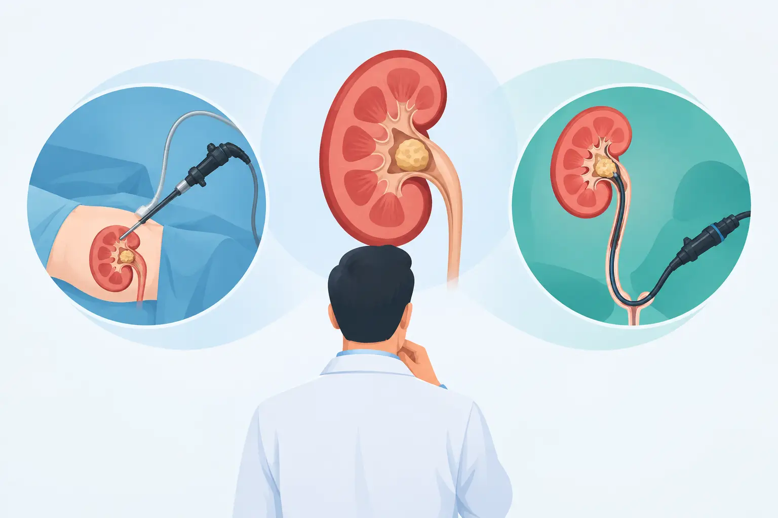
Imaging methods such as computed tomography help evaluate the entire urinary system, including the kidneys and ureters. This is because, in some patients with a bladder tumor, additional findings in the upper urinary tract should also be investigated.
The day of surgery and the procedure
TURBT is mostly performed under spinal anesthesia or, depending on the patient's condition, general anesthesia. The surgeon advances the camera system through the urethra into the bladder and systematically evaluates the entire inner surface of the bladder. The location, appearance, number and size of the tumor are recorded.
The tumor tissue is then removed in a controlled way. With large tumors, the procedure may take longer; additional measures may be needed to control bleeding. Correctly separating the removed tissues is one of the technical details that affect the quality of the pathological evaluation.
At the end of the procedure, a temporary catheter may be placed in the bladder. The catheter helps urine drain easily and allows any bleeding to be monitored. In some patients, the bladder may need to be irrigated with fluid. The length of hospital stay varies according to the size of the tumor, bleeding during the procedure, accompanying illnesses and the color of the urine. While the patient may be discharged quickly with small and limited tumors, close monitoring may be needed after more extensive resections.
What to expect after surgery
In the first days, slight pinkness in the urine, frequent urination, a burning sensation or a sudden urge to urinate may occur. These are temporary in most patients. However, in case of bright red bleeding with clots, a blocked catheter, fever, severe pain, inability to urinate or increasing fatigue, the doctor should be contacted without delay.
During recovery, it is important to drink enough fluids, avoid heavy exercise for the period recommended by the doctor and take prescribed medications regularly. When to restart medications that may increase the risk of bleeding is determined according to the plan made before the procedure. Each patient's pace of recovery and time to return to daily life are not the same.
The pathology result determines the direction of treatment
After minimally invasive surgery, the most awaited information is the pathology result. The result evaluates whether the tumor is low or high grade, which layer of the bladder wall it has reached, whether muscle tissue is present in the sample and whether accompanying flat carcinoma in situ is present.
In disease that has not progressed into the muscle layer, the risk of recurrence and progression is classified as low, intermediate or high. For some low-risk tumors, regular cystoscopy follow-up may be sufficient. In intermediate- and high-risk patients, drug treatments given into the bladder come into consideration. A single early dose of a chemotherapeutic agent or BCG immunotherapy given according to specific schedules can be planned based on the pathology and risk group.
BCG treatment is not necessary for every patient and is not applied the same way in every case. It is an important option aimed at preserving the bladder, especially in high-risk non-muscle-invasive bladder cancers. Even so, some tumors may recur or progress. For this reason, the success of treatment is evaluated not only through surgery but through regular and disciplined follow-up.
Why is regular cystoscopy follow-up necessary?
Bladder cancer is a disease that can tend to recur, even at a non-muscle-invasive stage. The fact that the tumor appears to have been completely removed does not mean a new focus will not form in the future. For this reason, cystoscopy is performed at set intervals; when necessary, urine cytology, imaging and additional tests are planned.
The frequency of follow-up is not the same for everyone. The follow-up plan for a patient with a low-risk, single, small tumor is different from the plan for a patient with a high-grade or recurrent tumor. The aim is not to create an unnecessary burden of procedures, but to catch any possible recurrence early.
Smoking is one of the strongest modifiable risk factors for bladder cancer. Even if surgery and intravesical treatments are successful, continuing to smoke can increase the risk of recurrence. For this reason, quitting smoking should be treated as a complementary part of the treatment plan.
Is the minimally invasive method always sufficient?
The most important advantage of minimally invasive surgery is that both diagnosis and, in many patients, initial treatment can be carried out without an abdominal incision. A shorter recovery time and the ability to preserve the bladder are also important benefits. However, the method is not a solution on its own for all stages of the disease.
In muscle-invasive bladder cancer or high-risk disease, more extensive treatment may be needed depending on the pathology result of the minimally invasive surgery. Radical cystectomy is an operation that can be performed with open, laparoscopic or robotic techniques. Which approach is suitable should be evaluated together with the patient's age, kidney function, other illnesses and life expectancy, as well as the characteristics of the cancer.
A bladder tumor diagnosis can cause anxiety; the safest way to reduce uncertainty is to create a clear treatment and follow-up plan based on the pathology result. As in the approach of Assoc. Prof. Dr. Uygar Miçooğulları, discussing in detail the limits of the method, the possible need for additional treatment and the follow-up steps before surgery strengthens the patient's decision-making process. Do not put off your questions; accurate staging and timely follow-up are the foundation of bladder cancer management.