What to do during a kidney stone attack is not just a matter of trying to suppress the pain. Although severe pain spreading from the lower back to the groin, testicle or lower abdomen sometimes passes on its own, it is necessary to assess how far the stone is blocking the urinary tract, whether there is an accompanying infection and whether kidney function has been affected. Especially for people experiencing pain of this severity for the first time, a medical assessment at the right time prevents unnecessary waiting and risky delays.
How do you recognize a kidney stone attack?
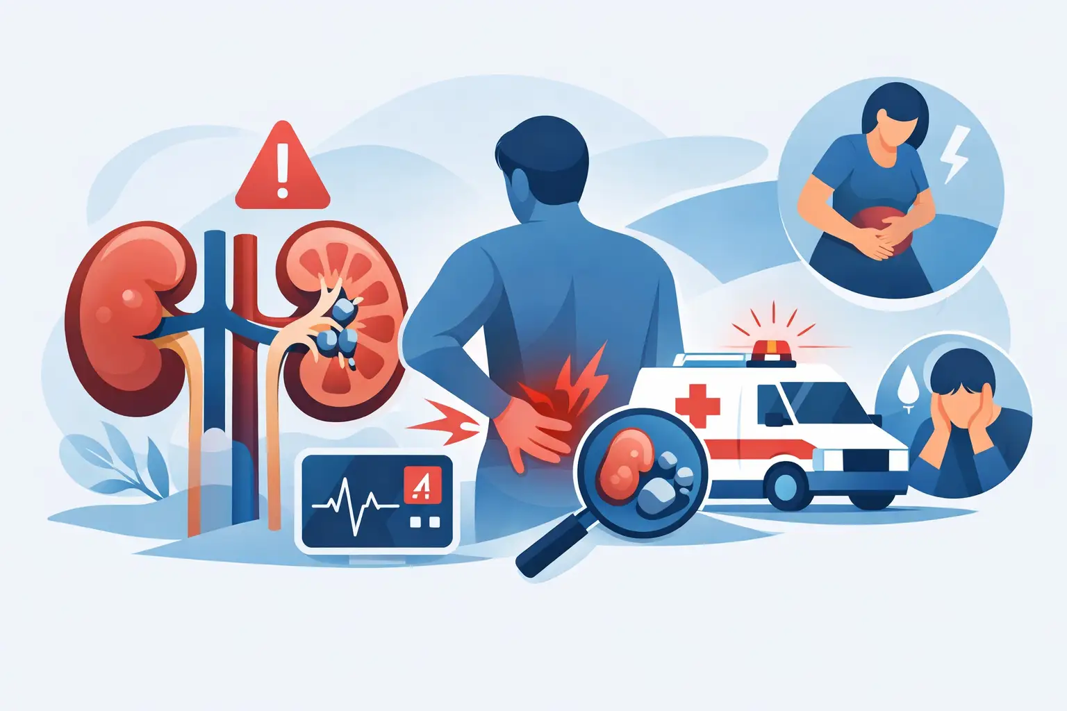
Kidney stone pain usually starts suddenly and intensifies in waves. The patient may find it hard to get into a comfortable position; the pain may start in the side of the lower back and spread to the lower abdomen, the groin or, in men, the testicle. Nausea, vomiting, burning on urination, a frequent urge to urinate and a pink-red change in urine color may also accompany the picture.
However, not every lower back pain is a stone attack. Musculoskeletal problems, bowel diseases, appendicitis, gallbladder disease and some vascular problems can cause similar complaints. For this reason, especially when the pain is unusually intense, persistent or affects your general condition, it is not right to make a diagnosis based on symptoms alone.
What to do in the first hours of a kidney stone attack
When the pain starts, the priority is to identify dangerous signs and to set up a safe assessment plan. If a doctor has previously recommended a painkiller for this situation, the prescribed dose and frequency can be followed. However, not every painkiller is suitable for people with stomach ulcers, kidney failure, blood thinner use, heart failure or certain blood pressure problems. Using someone else's medication or taking over-the-counter drugs one after another can be risky.
There is also a common mistake about drinking water. Trying to “flush the stone out with lots of water” is not always the right approach during severe pain and vomiting. Fluids can be taken in small sips to avoid dehydration; however, drinking excessive amounts of water can increase pressure and pain in a blocked urinary tract. If fluids cannot be kept down because of vomiting, you should go to a healthcare facility rather than forcing fluids by mouth.
If possible, note the time the pain started, any change in urine color, whether there is fever and the medications taken. If a stone has passed, it can be kept in a clean container. Stone analysis can guide the dietary and metabolic assessment aimed at reducing the formation of new stones.
Situations in which you should not wait at home
A stone attack may require emergency intervention when it occurs together with infection and urinary tract obstruction. If any of the following signs are present, go to the emergency department without delay:
- Fever, shivering, chills or marked weakness
- Severe pain that cannot be controlled despite painkillers
- Persistent vomiting, inability to take fluids or a feeling of faintness
- Inability to urinate or a marked decrease in urine output
- A known single kidney, kidney failure or a history of kidney transplant
- Severe flank pain developing during pregnancy
In a stone patient with fever, the aim is not just to relieve the pain. Infection of a blocked kidney can turn into a serious infection that spreads to the bloodstream. In this situation, antibiotics, urgent drainage of the urinary tract with a stent or nephrostomy and hospital monitoring may be needed.
Why is the diagnosis confirmed with imaging?
The size of the stone, its level and the dilation it causes in the kidney directly affect the treatment decision. A urine test provides information about signs of infection and bleeding, while blood tests help assess kidney function and markers of infection.
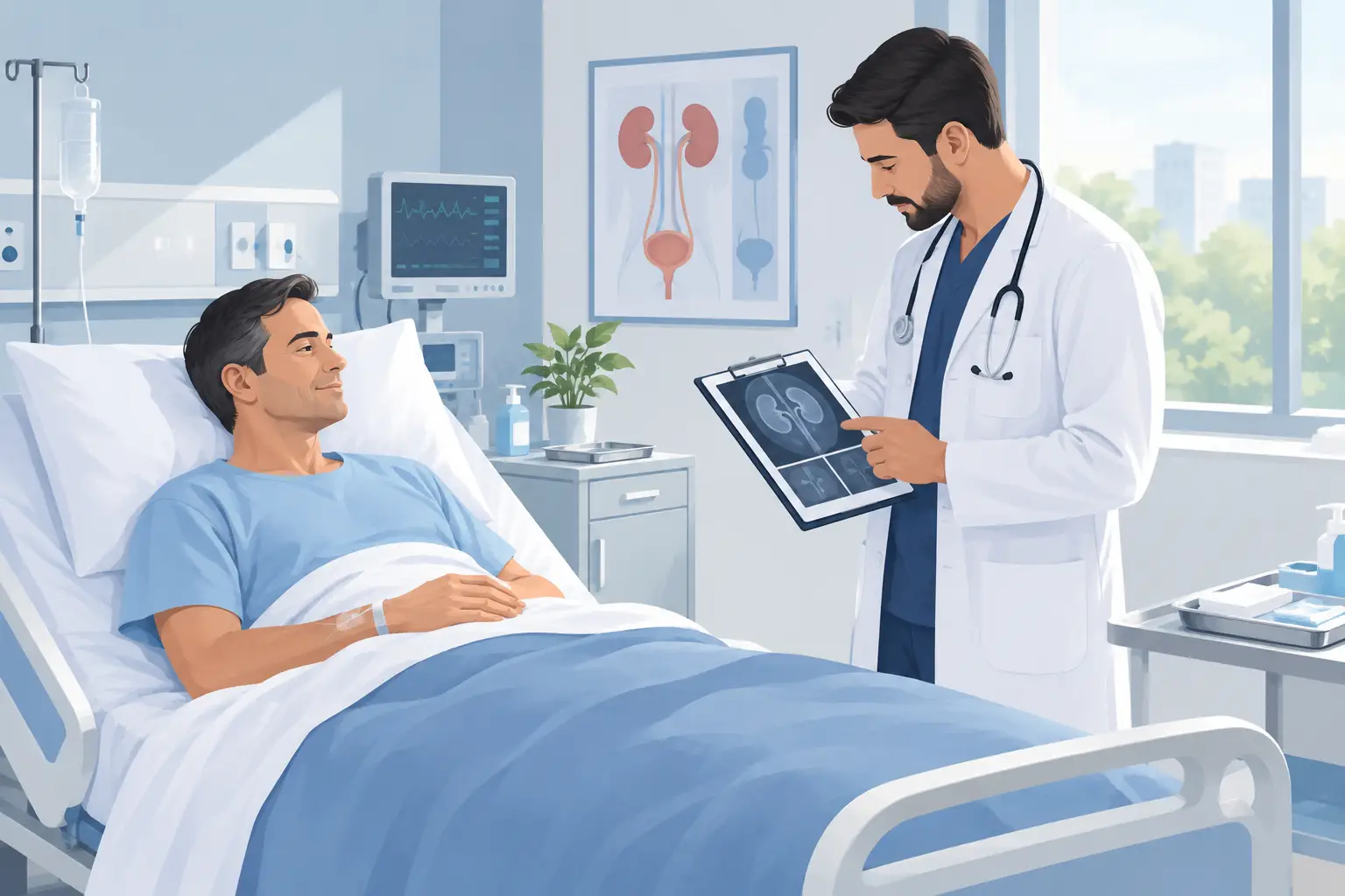
Ultrasonography is a valuable, radiation-free method for showing whether the kidney is dilated. However, small stones in the ureter in particular may not always be seen clearly on ultrasound. When needed, the location, size and density of the stone are determined in more detail with low-dose stone-protocol computed tomography. This assessment is important for distinguishing between the likelihood that the stone will pass on its own and the need for intervention.
Does every kidney stone require surgery?
No. Many small, favorably located ureteral stones can pass on their own with pain control and close follow-up. In some patients, medication that may help relax the ureteral muscles can be planned. However, these treatments are not suitable for every patient; blood pressure, other medications and the location of the stone must be taken into account.
When the decision is to wait, follow-up should not be neglected. A decrease in pain does not necessarily mean the stone has passed. A silent, ongoing blockage can harm kidney function. For this reason, follow-up imaging should be done within the time recommended by the urologist.
Intervention comes into consideration if the stone is large, the pain keeps recurring, it causes marked dilation of the kidney, there is a risk of infection or it does not pass within a reasonable time. In modern urology, minimally invasive methods come to the fore depending on the location and size of the stone. With ureteroscopy (URS), a stone in the ureter can be reached with thin endoscopic instruments. For stones inside the kidney, flexible ureteroscopy and laser treatment, that is, the RIRS method, may be considered; for larger or complex stones, percutaneous nephrolithotomy (PCNL) options may be considered.
These methods are not chosen on the basis of a single criterion. The diameter and hardness of the stone, its position in the kidney, the patient's anatomy, accompanying conditions and previous stone treatments are considered together. The aim is to clear the stone effectively with the least invasive method possible and to protect the kidney.
Reducing stone recurrence after the attack has passed
Passing the first stone does not mean that the stone disease is over. Especially in people with a history of recurrent stones, the cause should be investigated with stone analysis, a 24-hour urine test and the necessary blood tests. Calcium, uric acid, oxalate and citrate levels and urine volume can form the basis of an individual prevention plan.
In most patients the general approach aims at adequate fluid intake spread throughout the day. However, in people with heart or kidney disease, the amount of fluid should be adjusted according to the doctor's advice. Reducing excessive salt intake, avoiding unnecessary high-dose vitamin C supplements and eating animal protein in moderation may also be helpful for some stone types. Cutting out calcium completely, however, is not right; normal dietary calcium intake can reduce oxalate absorption from the gut in some people.
The treatment and prevention plan should be based on the type of stone, not on general advice found online. For example, recommendations for uric acid stones and calcium oxalate stones may not be the same. The approach of Assoc. Prof. Dr. Uygar Miçooğulları likewise aims not only to remove the stone but also to assess the personal factors that affect the risk of recurrence.
Even if severe pain eases, rather than ignoring a stone attack, planning a urological assessment that clarifies the stone's effect on the kidney is the safest step. Accurate diagnosis and timely follow-up both prevent unnecessary intervention and ensure that, when needed, kidney-protecting treatment is reached without delay.